PriCells - normal human umbilical vein primary endothelial cell culture
First, experimental reagents
1. Medium: PriCells Medium + 10% FBS + 1% P/S + PriCells Supplem e nt
2 , cryopreservation solution: PriCells Medium + 20% FBS + 10% DMSO
3 , washing solution: 1 × PBS ( pH 7.4  ) + 1% P/S
4 , staining solution: 0.4% Trypan Blue Â
5 , digestive juice: PriCells Isolation of Primary Cell Kit
6. Detection reagent: anti-human factor VIII antibody, fluorescently labeled secondary antibody, ethanol-acetone mixture ( 1 : 1 )
Second, the experimental equipment
1 , Petri dish
2 , culture bottle
3 , direct cut and eye scissors
4 , ophthalmology and hemostats
5 , 10ml syringe
6 , glass dropper
7 , beaker
8 , 15ml centrifuge tube
Third, the experimental process
Material
↓
Wash the blood stains outside the umbilical cord and cut the tape to a length of about 15 cm .
↓
PBS lavage to remove congestion
↓
Push the air and drain the remaining 1 × PBS ( pH 7.4  )
↓
Syringe perfusion of digestive juice ( PriCells ), use the hemostatic forceps to cut off when the digestive juice flows out at the other end, continue to perfuse the digestive juice to the vascular filling, cut off the end with another hemostatic forceps, and place it in antibiotic 1 × PBS ( pH 7.4  ) in the beaker
↓
Place the beaker at 37 ° C , 15-20 min water bath, digest at constant temperature, shake at 2-3 min intervals
↓
Collect the digestive juice, wash the vein 3-4 times with medium , 3ml/ time, then add the digestion stop solution ( PriCells )
↓
Centrifugation, 1000rmp, 10min, discard the supernatant
↓
Resuspend, count cells
↓
Inoculation density is 5 × 10 5 /ml
↓
Culture at 37 ° C , 5% CO 2
Fourth, experimental operation
1. The culture bottle is pre-coated. The flask was pre-packaged one day before the test, placed in an ultra-static table or dried in an oven at 45 ° C (remember to keep it sterile), placed in a refrigerator at 4 ° C , and set aside.
2 , material: normal delivery of fetal umbilical cord, length 15-20cm .
3, pretreated material: acquired into the vessel 1 × PBS containing 1% P / S in (pH 7.4  ) Were washed repeatedly to remove the external umbilical cord blood, after repeated washing solution by syringe flushing live umbilical venous filling, into the blood vessel without blood, the washing solution clarifies.
4 , digestive juice: use the syringe to focus on the air into the blood vessel, discharge the residual washing liquid, inject the digestive juice from the opening of one end of the blood vessel , and when the other open end has the digestive juice flowing out, the blood vessel can be ligated with a hemostatic forceps, and the digestive juice is continuously infused. Until the blood vessels were filled, the blood vessels were also blocked with a hemostatic forceps and digested in a 37 ° C water bath for 30 min in a beaker containing sterile PBS .
5. Stop the digestion: remove the hemostat, let the enzyme solution flow into the pre-prepared culture dish, add the digestion stop solution according to 1:1, stop the enzyme reaction, and wash the blood vessel with the medium for 3 times.
6. Collect the resuspended cells: collect the digested solution after the suspension in a centrifuge tube , centrifuge at 1000 rpm for 10 min , discard the supernatant, resuspend the culture medium, and stain the cells.
7. Culture cell density: Adjust the cell density to inoculate the flask at 5 × 10 5 /ml .
8. Culture: Place in a 37 ° C , 5% CO 2 incubator.
V. Cell identification
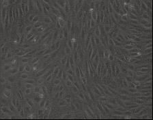
1. Microscopic identification: Under the phase contrast microscope, the cells are fusiform, and the cell density is increased, and the cobblestone-like mosaic arrangement is arranged, which has the characteristics of contact inhibition. The cells have good light transmission.
2 , immunohistochemical identification: using factor VIII-related antigen detection (or identification of endothelial cells using CD31 and CD33 immunofluorescence staining).
3 , cell climbing. The washed coverslips were placed in a 6- well culture plate, and the cells were seeded at 3 × 10 4 cells / well. At 48 hours, the cells were overgrown and removed with forceps.
Cover the cells with spare covers.
4. Cell fixation: The cells were washed with PBS , then placed in a mixture of ethanol and acetone, fixed for 10 min , and naturally dried in the air.
5. Specific antibodies: Dilute the antibody according to the dilution requirements, add the antibody, and incubate at 37 °C for 60 min .
6 , washing: 1 × PBS ( pH 7.4  ) Wash 3 times × 15 minutes and dry.
7. Labeled antibody: Add fluorescently labeled antibody and incubate at 37 °C for 30 min .
Washing: 1 × PBS ( pH 7.4  ) Wash 3 times × 15 minutes and dry.
8 , cover: sealing tablets.
9. Microscopic examination: Under the fluorescence microscope, the endothelial cell encapsulation showed strong yellow-green fluorescence, especially around the nucleus.
Six, matters needing attention
1. Select the material to select the umbilical cord with rounded and full distortion. No blood clots are blocked in the umbilical cord blood vessels, and the length of the umbilical cord is more than 15cm . The material collection process is aseptic as possible. If it is far away from the laboratory, the obtained umbilical cord can be washed with PBS containing antibiotics to remove residual blood, then immersed in the washing liquid, and the low temperature ice box is brought back to the laboratory.
2 , pay attention to eliminate residual blood in the blood vessels as much as possible, to avoid the influence of blood cells and certain serum components on endothelial cell adhesion.
3 , endothelial cell isolation damage seriously affects the growth of endothelial cells, so the enzyme digestion concentration and digestion time should be strictly controlled.
Â
4. Strictly control the culture conditions of endothelial cells. Including the quality and concentration of cell culture fluids (culture medium, growth factors, serum, antibiotics).
5 , Regarding the culture of the bottle or not, there is literature research showing that there is no particularly large difference between the coated and uncoated. In the experiment, it is possible to consider whether or not to coat.
6. The inoculation amount of the subculture cells was 5 × 10 4 ( 25 cm 2 flask), and the cell doubling time was 48 hours. Cell subculture is best for 5-8 generations, the number of passages is increased, the cell volume is increased, the gap between cells is increased, the cells are firmly attached, and the passage time is prolonged.
Seven, PriCells cell picture
|
You are always welcome to visit our company and taste our products. Contact: MS. Sunny Wang |
|
| Product Description | |
| Name | Canned Tuna |
| Flavor | Brine, Oil, Salad, Chili |
| Type | Shred, Flake, Chunk, Solid |
| Certificates | EU, FDA, BRC, HALAL,HACCP,KOSHER |
| Net weight | 140g, 160g, 170g, 185g, 200g, 1kg, 1.88k. |
| Brand | Our brand or OEM, ODM |
| Shelf life | 3/4 Years |
| MOQ | 1X20'FCL |
| Payment terms | T/T, L/C |
| Delivery time | 25 days after label artwork confirmed and advance payment done. |
| Packing | normal lid or easy open,paper label or lithio can, paper carton or shrinked by tray |
| EU NO. | 3302/01034 |
| RUSSIA NO. | 3302/01034 |
| Shipping docs | Commercial Invoice |
| Packing List | |
| Bill of Lading | |
| Certificate Of Origin/ Form A | |
| Health Certificate | |
| Veterinary Certificate | |
| Catching certificate | |
| Or as per customer`s request | |
Canned Tuna
Canned Tuna,Tuna Packets,White Meat Tuna,Canned Tuna In Sunflower Oil
Tropical Food Manufacturing (Ningbo) Co., Ltd. , https://www.tropical-food.com