Microglia are innate immune cells in the brain and play an important role in damage repair and anti-microbial invasion. However, under certain pathological conditions, microglia can exacerbate the inflammatory response and cause neurodegenerative changes. Therefore, targeted delivery of bioactive substances to microglia will help elucidate its mechanism of action and clarify its regulatory role in disease treatment. The Li Gan team at the University of California, San Francisco's Gladstone Institute for Neurological Diseases found that Quantum dots (QDs) were selected for targeting small glial cells in vitro and in vivo.
S Sakura Minami et al. isolated cerebral cortex tissue from neonatal rats in vitro, mixed cell culture, and then incubated with quantum dots. The results showed that quantum dots were selectively taken up by microglia, while neuronal cells and astrocytes did not acquire quantum dots (Fig. 1A-D). Further experiments suggest that the uptake of quantum dots by microglia is not affected by surface chemical modification. Different surface-modified quantum dots such as QD655-PEG, QD655-PEG-Strep and QD655-Carboxy are taken up by microglia. At the same time, quantum dots of different particle sizes (QD525, 605, 655 and 705) were taken up, and QD655 was the most efficient to enter cells (Fig. 1E-F). After the quantum dots enter the microglia, they do not induce cytotoxicity and do not affect the release of inflammatory factors by microglia. Quantum dots were injected into the hippocampus of mice, and fluorescence imaging showed that the quantum dots were distributed in most areas of the hippocampus and were mainly localized to microglia (Fig. 2). The quantum dot fluorescence signal is strong and has no significant quenching and continues until 1 month after injection. The quantum dots are coupled to the cytotoxin, saporin, which can target microglia and cause death, thereby significantly improving neuronal activity in a neurodegenerative disease model. In conclusion, this study provides a new research method for the specific labeling and imaging of microglia, and is of great significance for the drug delivery of small glial cells and the treatment of nervous system diseases.

Figure 1 Quantum dots are selectively taken up by microglia in primary mixed culture of rat cortical tissue.
(A) Microglia are specifically labeled with CD11b and Iba-1 antibodies (green), quantum dots (red) are mainly taken up by microglia; (B) quantum dots (red) are not astrocytes ( Ingested by the GFAP antibody as green);
(C) quantum dots (red) are not taken up by neuronal cells (marked green by MAP2 antibodies);
(D) As the incubation time increases, the fluorescence intensity of quantum dots taken up by microglia gradually increases;
(E) No quantum dots were detected in apoptotic or necrotic cells labeled with Annexin V/SyTox green (green);
(F) Quantum dots with different surface modifications are taken up by microglia.
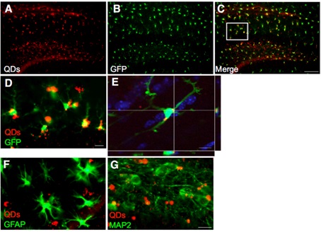
Figure 2 Quantum dots are selectively taken up by microglia in a body experiment.
(A)-(D) Stereotactic injection of quantum dots into CX3CR+/- mice, wherein the microglia have GFP expression.
(A) Distribution of quantum dots in the hippocampus;
(B) Microglia with GFP expression in hippocampus;
(C) a combination of A and B; (D) a partial enlargement of the C diagram;
(E) Confocal images of a single quantum dot plane taken up by hippocampal microglia;
(F) quantum dots (red) are not taken up by astrocytes (marked green by GFAP antibodies);
(G) Quantum dots (red) were not taken up by neuronal cells (marked green by MAP2 antibody).
Source of the document:
Minami SS, Sun B, Popat K, Kauppinen T, Pleiss M, Zhou Y, Ward ME, Floreancig P, Mucke L, Desai T, Gan L. Selective targeting of microglia by quantum dots. J Neuroinflammation. 2012;9:22.
1 ml sterile hypodermic syringe,2ml syringe,dispensing syringe,auto disable safety syringe
Shandong Zhushi Pharmaceutical Group Co.,LTD , https://www.sdzs-medical.com